 http://www.eorthopod.com/
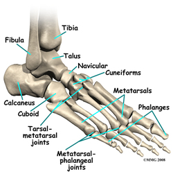
The foot and ankle are comprised of many bones. The lower leg bones include the tibia and fibula. The tarsal bones include the calcaneous, talus, navicular, cuboid, and three cuneiforms. There are five metatarsals in the mid foot that extend to the phalanges. The phalanges are also known as your toes.
There are also several joints that comprise the foot and ankle. The talocrural joint is made up of the tibia, fibula, and talus. This is where many of the motions of the ankle occur and create stability between the foot and lower leg. The subtalar joint is between the talus and the calcaneous. The tarsalmetatarsal joint joins the tarsal bones with the metatarsals. The metatarsalphalangeal joint connects the midfoot of the metatarsals to the toes, also known as the phalanges. All of these joints are important for correct movements to occur in the foot and ankle for walking, running, jumping, and lateral movements.
 http://www.orlandparkorthopedics.com
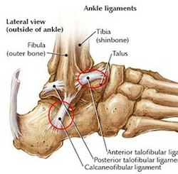
Lateral ankle ligaments include the anterior talofibular ligament, calcaneofibular ligament, and posterior talofibular ligament. These ligaments get their names from the bones they connect together including the talus, fibula, and calcaneous. They are especially important in stability of the lateral ankle and can become easily damaged with an inversion ankle sprain.
 https://classconnection.s3.amazonaws.com
Medial ankle ligaments include the posterior tibiotalar ligament, tibiocalcaneal ligament, anterior tibiotalar ligament, and tibionavicular ligament. Together these ligaments are commonly known as the deltoid ligament because they are a broad, thick band that mesh together. They also get their names from the bones they attach to including the tibia, talus, calcaneous, and navicular. Whiile it is difficult to injury this strong ligament, it is possible with an eversion ankle sprain.
 http://www.physioadvisor.com.au
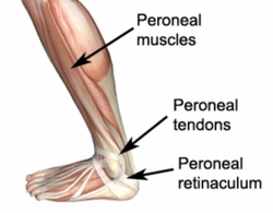
Lateral ankle muscles include the peroneal muscles including peroneal longus and peroneal brevis. The peroneal longus and brevis are responsible for eversion and plantarflexion of the foot.
 http://www.aafp.org/afp/2004/0715/afp20040715p332-f1.jpg
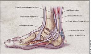
Medial ankle muscles include the flexor hallucis longus, posterior tibialis, and flexor digitorum longus while the anterior muscles include the anterior tibialis, extensor hallucis longus, and extensor digitorum longus. The flexor hallucis longus flexes the great toe and plantarflexs and inverts the foot. The posterior tibialis plantarflexs and inverts the foot. The flexor digitorum longus flexs the second through fifth toes while inverting and plantarflexing the foot. The anterior tibialis is resposible for dorsiflexion and inversion. The extensor hallucis longer lies on the anterior side of the ankle and is responsible for dorsiflexion and extension of the great toe. The extensor digitorum longus also dorsiflexs the foot and extends the second through fifth phalanges.
 https://classconnection.s3.amazonaws.com
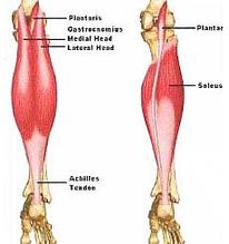
The posterior muscles include the gastrocnemius and soleus. Together these two muscles are known as the triceps surae group. They are both responsible for plantarflexion of the foot. The gastrocnemius also flexes the knee. They both insert into the Achilles tendon on the calcaneous on the foot.
Information can be found from....
Starkey, C., Brown, S., & Ryan, J. (2010). Ankle and Leg Pathologies. In Examination of Orthopedic and Athletic Injuries (Ed. 3. ed., pp. 221-285). Philadelphia, Pennslyvania: F.A. Davis.
Starkey, C., Brown, S., & Ryan, J. (2010). Ankle and Leg Pathologies. In Examination of Orthopedic and Athletic Injuries (Ed. 3. ed., pp. 221-285). Philadelphia, Pennslyvania: F.A. Davis.
